by Dr. Stephen Pelsue
Wed, Sep 17th, 2025 8:48 am
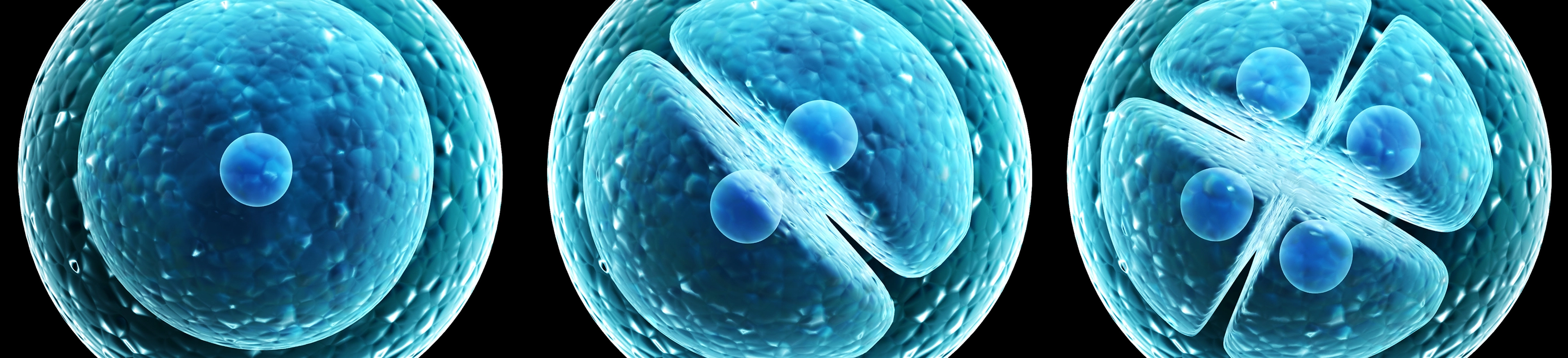
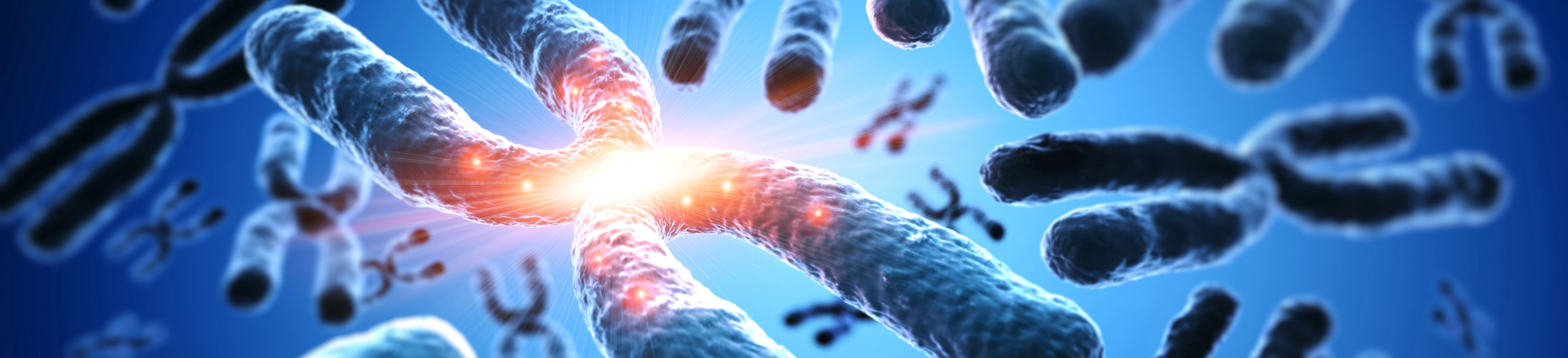
When cells divide, they must ensure that each daughter cell receives an identical set of chromosomes. To achieve this, each chromosome is first duplicated, forming two identical sister chromatids that are connected at a central point called the centromere. During cell division, these duplicated chromosomes align at the center of the cell. Spindle fibers, made of microtubules, attach to the chromosomes at the centromeres onto protein structures called kinetochores, and also anchor to opposite sides of the cell. The spindle fibers then pull the duplicated chromatids apart, directing one copy to each end of the cell. This process, known as chromosome segregation, ensures that both daughter cells end up with the same chromosome content.
Proper spindle attachment is therefore critical to ensure accurate and balanced chromosome distribution. Consequently, this process must be tightly controlled so that each chromosome is attached at only one specific site.
The centromere is mostly made up of short, repeating DNA sequences of around 150 base pairs (bp) long. These short repeats are then organized into larger repeating units of about 2kb in size. These larger units are repeated over and over again to form what's called the alpha-satellite array, the main structure of the centromere. Altogether, a centromere can span hundreds of thousands to millions of base pairs. Because it is made entirely of highly repetitive DNA, it's been very difficult to sequence and study accurately using traditional genome sequencing methods.
A recent effort by one group used both long-read sequencing technology (nanopore sequencing) and short single molecule sequencing to look in detail at human centromeres. What they found was quite interesting: the variation of centromere size and sequence was much greater than expected. As this structure is required for a key function, the expectation was that both the sequence and the size would be highly conserved. The fact that there are a high-degree of mutation and a rapid evolutionary rate may give some insight into chromosome abnormalities.
It is unknown if mutations lead to altered function, but the size of the centromere is likely to have a significant impact: too small and the kinetochore structure is unlikely to form properly; too large and multiple attachments can take place that could lead to breakage of the chromosome. With new technology now enabling the study of centromeres, researchers can explore the extent of their variation and investigate how their sequence and structure contribute to aneuploidy, aging, cancer, and normal development.